Header Graphic
Dr. Paul Degenaer
Therapeutic Optometrist
Retinopathy

Diabetic Eye Disease

If you have diabetes mellitus, your body does not use and store sugar properly.  High blood sugar levels can damage blood vessels in the retina, the nerve layer at the back of the eye that senses light and helps to send images to the brain.   The damage to retinal vessels is referred to as diabetic retinopathy.

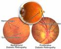
Type of diabetic retinopathy

There are two types of diabetic retinopathy:  nonproliferative diabetic retinopathy (NPDR) and proliferative diabetic retinopathy (PDR).

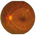
NPDR, commonly know as "background retinopathy," is an early stage of diabetic retinopathy.  In this stage, tiny blood vessels with the retina leak blood or fluid.  The leaking fluid causes the retina to swell or to form deposits called exudates.  Many people with diabetes have mild NPDR, which usually does not affect their vision.  When vision is affected it is the result of macular edema and / or macular ischemia.

Macular edema is swelling, or thickening, of the macula, a small area in the center of the retina that allows us to see fine details clearly.  The swelling is caused by fluid leaking from retina blood vessels.  It is the most common cause of visual loss in diabetes.  Vision loss may by mild to severe, but even in the worst cases, peripheral vision continues to function.

Macular ischemia occurs when small blood vessels (capillaries) close.  Vision blurs because the macula no longer receives sufficient blood supply to work properly.  
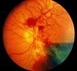
PDR is present when abnormal new vessels (neovascularization) begin growing on the surface of the retina or optic nerve.  The main cause of PDR is widespread closure of retinal blood vessels, preventing adequate blood flow.  The retina responds by growing new blood vessels in an attempt to supply blood to the area where the original vessels closed.

Unfortunately, the new, abnormal blood vessels do not resupply the retina with normal blood flow.  The new vessels are often accompanied by scar tissue that may cause wrinkling or detachment of the retina.

PDR may cause more severe vision loss than NPDR because it can affect both central and peripheral vision.

Proliferative diabetic retinopathy causes visual loss in the following ways:

Vitreous hemorrhage: The fragile new vessels may bleed into the vitreous, a clear jelly-like substance that fills the center of the eye.  If the vitreous hemorrhage is small, a person might see only a few new dark floaters.  A very large hemorrhage might block out all vision.

It may take days, months, or even years to resorb the blood, depending on the amount of blood present.  If the eye does not clear the vitreous blood adequately within a reasonable time, vitrectomy surgery may be recommended.  Vitreous hemorrhage alone does not cause permanent vision loss.  When the blood clears, visual acuity may return to its former level unless the macula is damaged.

Traction Retinal Detachment: When PDR is present, scar tissue associated with neovascularization can shrink, wrinkling and pulling the retina from its normal position.  Macular wrinkling can cause visual distortion.  More severe vision loss can occur if the macula or large areas of the retina are detached.

Neovascular Glaucoma: Occasionally, extensive retinal vessel closure will cause new, abnormal blood vessels to grow on the iris (colored part of the eye) and block the normal flow of fluid out of the eye.  Pressure in the eye builds up, resulting in neovascular glaucoma, a severe eye disease that causes damage to the optic nerve.

How is diabetic retinopathy diagnosed?

A medical eye examination is the only way to find changes inside your eye.  An ophthalmologist optometrist can often diagnose and treat serious retinopathy before you are aware of any vision problems.  The ophthalmologist dilates your pupil and looks inside of the eye with an ophthalmoscope.

If your ophthalmologist or optometrist finds diabetic retinopathy, he or she may order color photographs of the retina or a special test called a fluorescein angiogram to find out if you need treatment.  In this test a dye is injected in your arm and photos or your eye are taken to detect where fluid is leaking.

How is diabetic retinopathy treated?

The best treatment is to prevent the development of retinopathy as much as possible.  Strict control of your blood sugar will significantly reduce the long-term risk of vision loss from diabetic retinopathy.  If high blood pressure and kidney problems are present, they need to be treated.

Laser surgery:  Laser surgery is often recommended for people with macular edema, PDR and neovascular glaucoma.
For macular edema, the laser is focused on the damaged retina near the macula to decrease the fluid leakage.  The main goal of treatment is to prevent further loss of vision.  It is uncommon for people who have blurred vision from macular edema to recover normal vision, although some may experience partial improvement.  A few people may see the laser spots near the center of their vision following treatment.  The spots usually fade with time, but may not disappear.

For PDR, the laser is focused on all parts of the retina except the macula.  This panretinal photocoagulation treatment causes abnormal new vessels to shrink and often prevents them from growing in the future.  It also decreases the chance that vitreous bleeding or retinal distortion will occur.

Multiple laser treatments over time are sometimes necessary.   Laser surgery does not cure diabetic retinopathy and does not always prevent loss of vision.

Vitrectomy:  In advanced PDR, the ophthalmologist may recommend a vitrectomy.  During this microsurgical procedure, which is performed in an operating room, the blood-filled vitreous is removed and replaced with clear solution.  The ophthalmologist may wait for several months to see if the blood clears on its own before performing a vitrectomy.

Vitrectomy often prevents further bleeding by removing the abnormal vessels that caused the bleeding.  If the retina is detached, it can be repaired during the vitrectomy surgery.  Surgery should usually be done early because macular distortion or traction retinal detachment will cause permanent visual loss.  The longer the macula is distorted or out of place, the more serious the vision loss will be.

Copyright 1998   American Academy of Ophthalmology


Contact Us                                                                           

Senior Sight Inc.                                                                                                                              

5001 McKinney Ranch Pkwy. Suite A                                     

McKinney, Texas 75070       

972-547-4200

Homepage

Informacion en Espanol              

Review from Solutionreach: "Efficient, fast and easy to understand process!"